

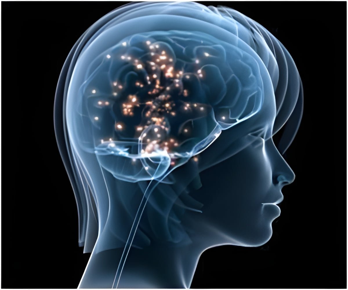
A world-first AI-assisted brain surgery has helped save the sight of a 48-year-old man undergoing removal of a pituitary tumor that was affecting his vision. The AI analyzed live surgical video while doctors operated.
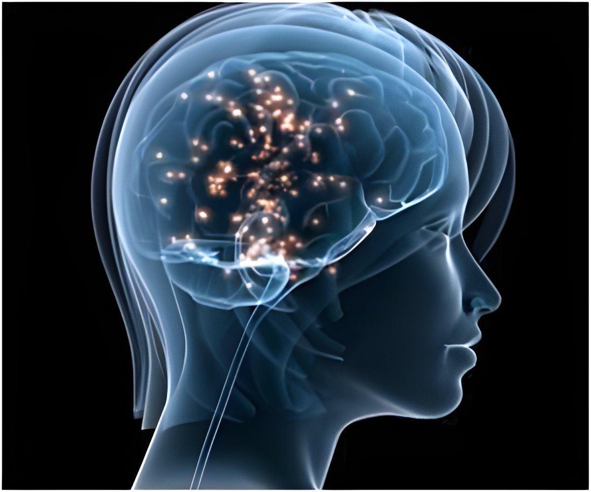
- World-first AI-assisted brain surgery used AI during a live human operation
- AI analyzed live endoscopic video to help identify critical anatomy
- The patient’s vision improved dramatically after removal of the pituitary tumor
A 48-year-old man has become the world’s first patient to undergo live AI-assisted neurosurgery, with artificial intelligence analyzing the surgical video in real time as doctors removed a pituitary tumor that was threatening his eyesight (1✔ ✔Trusted Source
First patient in live AI assisted sight-saving brain surgery
).
The pioneering operation was performed at the National Hospital for Neurology and Neurosurgery (NHNN), UCLH, as part of a clinical trial using AI technology developed at University College London (UCL) and supported by the National Institute for Health and Care Research (NIHR).
The patient, Rhys Hibbert from Bedfordshire, had developed worsening vision problems after being diagnosed with a pituitary tumor. During the operation, the AI system examined the live endoscopic camera feed and helped the surgical team recognize critical structures around the tumor.
Advertisement
Why Was This Brain Surgery a World First?
The breakthrough was not simply that AI was involved in brain surgery. Researchers had already developed artificial intelligence systems capable of analyzing surgical videos and recognizing anatomy.
What made the procedure different was that the technology was being used live during the operation, giving the surgical team additional information while they were operating.
The AI system analyzed the surgeon’s real-time endoscopic view, rather than relying only on scans obtained before surgery.
It was designed to help identify important anatomical structures and highlight areas that could be risky while the surgeon removed the tumor. This is particularly important in pituitary surgery, where the tumor is located close to delicate nerves and blood vessels.
Earlier research had already provided evidence that AI could improve anatomical recognition during pituitary surgery. A study published in npj Digital Medicine tested AI assistance among 24 participants, ranging from medical students to experienced surgeons. The study found that: (2✔ ✔Trusted Source
Artificial intelligence assisted operative anatomy recognition in endoscopic pituitary surgery
- AI helped doctors identify the tumor area more accurately, with accuracy improving from 70.7% to 77.5%.
- Medical students benefited the most, with their accuracy rising from 66.2% to 78.9%.
- AI also helped reduce mistakes in identifying important areas during surgery.
- This could be important because a wrong identification could damage nearby blood vessels or nerves.
However, that study was still preclinical. The researchers tested the system on still images rather than using it to assist a surgeon during a live operation.
They noted that further work was needed to develop real-time video capabilities, an appropriate display interface, and measures of safety, effectiveness, and efficiency before the technology could become an intraoperative decision-support tool.
Why Is a Pituitary Tumor So Dangerous for Vision?
The pituitary gland is extremely small, but its location makes surgery particularly delicate.
It sits at the base of the brain, close to the optic nerves and optic chiasm, the region where the optic nerves partially cross. These nerves carry visual information from the eyes to the brain.
A pituitary tumor can press against the optic nerves or optic chiasm. This can interfere with the transmission of visual information and produce symptoms such as (3✔ ✔Trusted Source
How pituitary tumours can affect your eyesight
- Blurred vision
- Double vision
- Reduced peripheral vision
- Loss of part of the visual field
Larger pituitary tumors, called macroadenomas, are more likely to cause visual problems because they have a greater chance of pressing against the optic nerves or optic chiasm.
The additional research on AI in pituitary surgery explains why this anatomy creates such a difficult surgical environment. Pituitary tumors can compress, distort, or surround nearby neurovascular structures, including the optic nerves and internal carotid arteries.
Surgeons commonly remove these tumors through an endoscopic endonasal trans sphenoidal approach, reaching the pituitary region through the nasal passages.
But identifying anatomy during the operation can be difficult. Tumors may distort normal structures, while previous surgery, radiotherapy, sinonasal disease, and natural anatomical differences can make orientation more challenging.
Conventional technology already helps surgeons. Endoscopes provide visual access, neuronavigation uses preoperative imaging to guide orientation, and selected procedures may use micro-Doppler or neurophysiological monitoring.
But each has limitations. For example, neuronavigation relies on preoperative imaging and may not fully account for changes in anatomy during surgery.
This is where real-time AI could potentially offer something different: analyzing what the surgeon is seeing at that exact moment.
Advertisement
How Did AI Actually Help the Surgeons?
During Rhys’s operation, AI watched the live video coming from the surgical camera and helped the team identify important structures around the tumour. This gave the surgeons an additional source of information while they were working in an area where nerves and blood vessels are very close together.
The technology was not designed to take over the operation. Instead, it was used as an extra set of eyes to help the surgeons recognize what they were seeing and decide where it was safer to operate.
This approach has been developed through several years of research into how AI can understand what happens during pituitary surgery. Researchers have trained AI systems to recognise:
- Different stages of the operation
- Surgical instruments
- Important structures in the surgical area
- How surgical instruments interact with tissue
A review describes computer vision as one of the most promising ways AI could eventually support surgeons during pituitary operations . These systems could provide useful information while surgery is happening, rather than analyzing the operation only afterwards.
The research behind these systems has been built using recordings of previous operations. One AI model was trained using 64 videos of endoscopic pituitary tumor surgeries, with 640 images taken from these operations to teach the system to recognize the surgical area.
Researchers have also developed systems specifically for real-time use. One example is PitSurgRT, which was designed to identify important structures during endoscopic pituitary surgery as the procedure is taking place.
These earlier developments helped pave the way for the live AI-assisted operation performed at UCLH. The technology can potentially help surgeons recognise important anatomy, follow surgical instruments and understand what is happening at different stages of the procedure.
The AI system used at UCLH runs on an NVIDIA Clara IGX platform, which is designed to support real-time AI applications in medical settings.
Most importantly, the surgeon remained in control throughout the operation. AI provided additional information to the surgical team, while the surgeons made the decisions and carried out the tumor removal.
Advertisement
What Happened to Rhys After the Operation?
Rhys’s pituitary tumor was discovered unexpectedly in December 2024 after he collapsed during a walk and experienced a seizure.
Medical investigations subsequently revealed an approximately 11-mm tumor in his pituitary gland. Initially, doctors managed his condition without surgery, but his symptoms later worsened.
Among the most serious problems was progressive visual impairment.
Rhys described losing peripheral vision to such an extent that he began using walking sticks after repeatedly tripping because he could not see the lower part of his visual field.
When surgery became necessary, Rhys decided not only to undergo the procedure but also to participate in the research.
He said he believed patients needed to take part in research if doctors were to learn and medicine was to advance. When he was asked whether he would become the first patient to undergo live AI-assisted surgery, he agreed.
The result was particularly encouraging (4✔ ✔Trusted Source
Artificial intelligence in pituitary surgery: the path to clinical solutions
After waking from surgery, Rhys said his vision had dramatically improved and that he could see the room clearly. Within approximately a week, he was walking independently without glasses or walking sticks.
He described the change as feeling like he had regained a 360-degree panoramic view of his surroundings.
Can AI Make Surgery Safer?
Research so far suggests that AI could improve safety at different stages of surgery, although it has not yet been proven to improve outcomes in every type of operation.
A review in Patient Safety in Surgery found promising results across studies, including AI systems that supported real-time decisions, recognized surgical instruments, helped analyze tissue, and detected complications earlier (5✔ ✔Trusted Source
Artificial intelligence and machine learning approaches for patient safety in complex surgery: a review
Some of the key findings include:
- During surgery: An AI warning system helped reduce the severity and duration of dangerously low blood pressure by about four times in one clinical trial.
- Recognizing surgical tools: AI was able to detect and count surgical instruments with 99.4% accuracy in one study.
- During brain surgery: An AI system analyzed brain tumor tissue in about 2.5 minutes, with 94.6% accuracy, providing rapid information to surgeons.
- After surgery: AI detected surgical-site infections up to 48 hours earlier than routine diagnosis and reduced the workload involved in monitoring patients.
- There are still limitations: Many studies have been conducted in single hospitals, used past patient data, or involved early-stage AI systems. More real-world research is needed to determine whether these technologies actually reduce complications and improve patient outcomes.
Overall, the research suggests that AI can support specific safety-related tasks, but it is still a tool to assist doctors and surgeons rather than replace them.
What Comes Next for AI in Brain Surgery?
The UCLH operation could be an early glimpse of a broader role for AI in pituitary care, from earlier diagnosis to real-time surgical assistance and monitoring after surgery.
Before surgery, AI could help identify pituitary disease earlier using medical records and imaging. During surgery, it could help surgeons recognize important anatomy and track instruments in real time.
After surgery, AI may eventually help predict complications, hormone problems and tumor recurrence.
However, AI still needs more testing before it can become a routine part of pituitary care. Researchers need larger and more diverse studies to assess its reliability, safety, and effectiveness, while also addressing concerns such as bias, data quality, and accountability.
For now, the goal is not to replace neurosurgeons but to give them better information and support during complex procedures. Rhys Hibbert’s successful operation is an important first step, but further research will determine whether this technology can consistently make surgery safer and improve outcomes for more patients.
Frequently Asked Questions
Q: What Made This a World-First AI-Assisted Brain Surgery?
A: The operation was the first reported human brain surgery in which AI analyzed live endoscopic surgical video to assist doctors during the procedure.
Q: How Did AI Assist During the Brain Surgery?
A: The AI analyzed the live surgical view and helped the surgical team recognize important anatomical structures around the pituitary tumor.
Q: What Type of Tumor Was Removed?
A: The surgeons removed a pituitary tumor that was affecting the patient’s vision by pressing on structures involved in sight.
Q: Did the Patient’s Sight Improve After Surgery?
A: Yes. The patient reported a dramatic improvement in his vision after the tumor was removed and was able to walk independently without glasses or walking sticks within about a week.
Q: Does AI Perform the Brain Surgery?
A: No. The surgeon remains in control. AI provides additional information and visual assistance rather than independently performing the operation.
Q: Could AI Make Brain Surgery Safer?
A: AI could potentially help surgeons recognize critical anatomy, track instruments and provide real-time guidance, but larger clinical studies are still needed to prove that it consistently improves patient safety and outcomes.
References:
- First patient in live AI assisted sight-saving brain surgery – (
https://www.uclh.nhs.uk/news/first-patient-live-ai-assisted-sight-saving-brain-surgery) - Artificial intelligence assisted operative anatomy recognition in endoscopic pituitary surgery – (https://www.nature.com/articles/s41746-024-01273-8)
- How pituitary tumours can affect your eyesight – (https://www.pituitary.org.uk/information/pituitary-tumours-and-eyesight/)
- Artificial intelligence in pituitary surgery: the path to clinical solutions – (https://journals.bioscientifica.com/erc/article/33/8/e260104/77942/Artificial-intelligence-in-pituitary-surgery-the)
- Artificial intelligence and machine learning approaches for patient safety in complex surgery: a review – (https://link.springer.com/article/10.1186/s13037-025-00458-8)
Source-Medindia